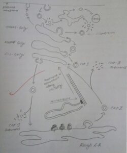
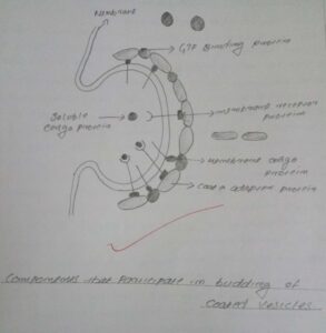
There are various small vesicles that transport proteins from one organelle to another. These vesicles bud from the membrane of particular parent organelle and fuse with the membrane of a particular “Target” (Destination) organelle.
They are critical for sorting of proteins newly made in the rough endoplasmic reticulum and also for proteins internalized from the cell surface.
There are three types of coated vesicles, each with a different type of protein coat and each formed by reversible polymerization of a distinct set of protein subunit under regulated conditions.
The Coated Vesicle Types are:
- Clathrin
- COP – I
- COP – II
The above three coat proteins are mainly involved in the vesicular traffic
The coats depolymerize into their subunits, After formation of vesicles by membrane budding. The depolymerized coat subunits are re-used to form additional vesicles.
Clathrin mediates transfer of vesicles that bud from the trans-Golgi network and the plasma membrane and that then fuse with late endosomes.
COP I mediates retrograde transport from the trans- to the medial- to the cis-Golgi, as well as from the cis-Golgi/cis-Golgi network to the rough ER. It may also mediate forward transfer of vesicles from the rough ER to the cis-Golgi network
COP II mediates transfer of vesicles from the rough ER to the cis-Golgi/cis-Golgi network; in some cases COP II-coated vesicles fuse with each other to form larger “ER-to-Golgi intermediate compartments” that are transported along a microtubule and eventually fuse to form the cis-Golgi.
Clathrin
Typical clathrin coated vesicles are 50 – 100 nm in diameter. The membrane bound coat vesicle consists of fibrous protein clathrin. Clathrin molecules have three limbed shape and are called Transkelion. Each limb contains one clathrin heavy chain and one clathrin light chain.
Even in the absence of membrane vesicles, clathrin transkelions can polymerize to form the cage like structure found around the coated vesicles. when clathrin polymerizes it forms a polygonal lattice with an intrinsic curvature.
Reference
Lodish H, Berk A, Zipursky SL, et al. Molecular Cell Biology. 4th edition. New York: W. H. Freeman
Cooper G M, The Cell: A Molecular Approach, ASM Press, USA
Got something to say about this post? Leave a comment…your comments are valuable for improving the posts.


